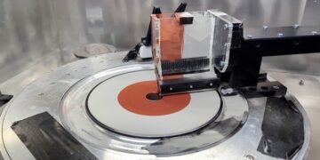
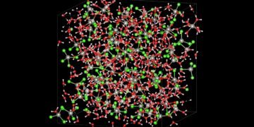
A group of scientists, made up of researchers from the Max Born Institute in Berlin and the Helmholtz-Zentrum Berlin in Germany and the Brookhaven National Laboratory at the Massachusetts Institute of Technology in the United States, have developed a revolutionary new method for producing high resolution, imaging of nanoscale structural changes using an intense X-ray source. This technique, which they call Coherent Correlation Imaging (CCI), allows creating sharp, detailed images without damaging the sample from excessive radiation. By using algorithms to detect patterns in unseen images, CCI opens the door to previously inaccessible information. The team demonstrated CCI in samples of thin magnetic layers, and have published their results in Nature.

The earth’s atmosphere is constantly moving forward and constantly changing. Even if something solid seems to be unchanging, these changes can lead to unusual things; An example is the zero transmission of electricity in high temperature superconductors. A transition is often called a phase transition, when a substance changes state, for example from solid to liquid during melting. Scientists also study changes in different systems, such as from a non-conductor to a conductor, from non-magnetic to magnetic, and changes in crystal structure. Many of these processes are used in technology and play an important role in the functioning of living organisms.
Problem: too much heat can damage the sample
However, studying these processes in detail is a difficult task, and taking a film of these changing processes is even more difficult. This is because the changes happen quickly and happen on the scale of nanometers – one millionth of a millimeter. Even the most advanced X-ray and electron microscopes cannot capture this rapid movement. The problem is rooted, as the principle of the picture shows: to capture a sharp picture of something, a level of light is required. To expand the object, i.e. “increase”, more light is needed.
More light is also needed when trying to capture fast and short exposures. Finally, increasing the resolution and reducing the exposure time lead to the point where the object will be damaged or even destroyed by the required light. This is exactly what science has reached in recent years: imaging using a free electron laser, the most powerful X-ray source available today, leads to the destruction of biological samples. Therefore, taking a movie of these random sequences with many frames is impossible.
A New Approach: Using Algorithms to Detect Patterns in Dimly Lit Images
An international team of scientists has found a solution to this problem. The main reason for their solution is to understand that the process of material change is not always random. Focusing on a small portion of the sample, the researchers found that some spatial patterns appeared repeatedly, but the time and timing of these patterns were unpredictable.
Scientists have developed a new non-destructive imaging technique called Coherent Correlation Imaging (CCI). To create a film, they take multiple snapshots of the sample in rapid succession while dimming the light enough to keep the sample. However, this results in individual images where the variable values in the view are not apparent.
However, the image still contains enough information to separate them into groups. To do this, the group must first develop a new algorithm that analyzes the relationship between images, hence the name of the system. The images within each group are very similar and are therefore likely to result from a specific change pattern.
Only when all the colors in the group are combined will there be a clear image of the sample. Scientists have now been able to restore the film and match each image with a clear image of the sample’s condition at the time.
One example: filming a “music section” with magnetic layers
Scientists developed this new method to solve a specific problem in the field of magnetism: the production of ferromagnetic material is important. These levels are divided into areas called sectors, in which the magnetization points up or down. A similar magnetic film is used in modern hard drives where two different types of sectors are assigned a “0” or “1” bit. So far, these types are believed to be stable. Or is it really true?
To answer this question, the team studied samples made of such magnetic layers at the National Synchrotron Light Source II on Long Island near New York, using the newly developed CCI method. In fact, the pattern does not change at room temperature. But at a slightly higher temperature of 37°C (98°F), the segments began to move in the wrong direction, twisting each other. The scientists observed this “horror section” for several hours. Finally, they created a map that shows the best place of border between the departments. This map and moving image provided insight into magnetic interactions with matter, supporting future applications in advanced computer processing.

New opportunities for material analysis in X-ray sources
The next goal of scientists is to use new imaging techniques and free electron lasers, such as the European XFEL in Hamburg, to better understand even fast processes at small length scales. They are convinced that this method will improve our understanding of the role of change in stochastic processes in modern materials and, therefore, find new ways to use them in a clear way.


