Imagine you have an MRI of the knee. This test measures the density of water droplets in your knee, with a resolution of about one cubic millimeter – which is good for determining if, for example, the meniscus is torn in the knee. But what if you were to study the structural data of a five nanometer cubic molecule, which is about ten billionths smaller than the best resolution current MRI scanners can produce? That is the purpose of Dr. Amit Finkler of the Department of Chemical and Biological Physics at the Weizmann Institute of Science. In a recent study, Finkler, doctoral student Dan Yudilevich and colleagues at the University of Stuttgart, Germany, succeeded in taking a big step in this direction, by showing a new method of using individual photo electrons. This method, which is still very small, may one day affect the imaging of different types of particles, which may change the development of medicine and the identification of quantum objects.
Current magnetic resonance imaging (MRI) techniques have been helpful in diagnosing a variety of diseases for decades, but while the technology has evolved over the years, some of the underlying problems have yet to be resolved. For example, the reading efficiency of MRI is very small, requiring a sample of hundreds of billions of fluids – if not more – to operate. A side effect of this inefficiency is that productivity is improved. For most research methods, the average is the best, but when you calculate many variables, some details are lost, which can obscure important processes that occur on a small scale.
Whether this is a problem or not depends on the question you are asking: for example, there is a lot of information that can be gleaned from a photograph of a crowd in a crowded football stadium, but a photograph cannot the best tool. will be used if someone wants to know more about the mole on the cheek of the person sitting in the third seat of the fourteenth row. If we want to collect more data on the mole, getting closer might be the way to go. Finkler and his collaborators actually provide molecular tracking. The use of such a tool can enable researchers to analyze the structure of important molecules and possibly open the way to new discoveries. In addition, there are times when a small “cloth” will be necessary for the work itself – such as during the early stages of drug development.

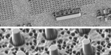
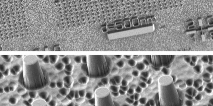
So how do we get an accurate MRI equation that can work with small samples – down to the individual? Finkler, Yudilevich and Stuttgart Dr. Rainer Stöhr and Andrej Denisenko have developed a method that can find the exact location of an electron. It is based on a rotating magnetic field that is close to the center of nitrogen space – a spot with the size of an atom in a special synthetic diamond, which is used as a quantum sensor. Because of its atomic size, this sensor is particularly sensitive to nearby changes; due to its quantum nature, it can identify whether one or more electrons are present, making it ideal for measuring the position of individual electrons with incredible precision.
“This new method,” says Finkler, “can be used to give a more detailed explanation to existing methods, with the goal of better understanding the Holy Trinity of processes, functions, and changes. ” For Finkler and his colleagues, this discovery is a very important step on the road to accurate nanoimaging, and they hope that in the future we can use this method to make different types of cells, which will measure look to be ready for their big plans.
