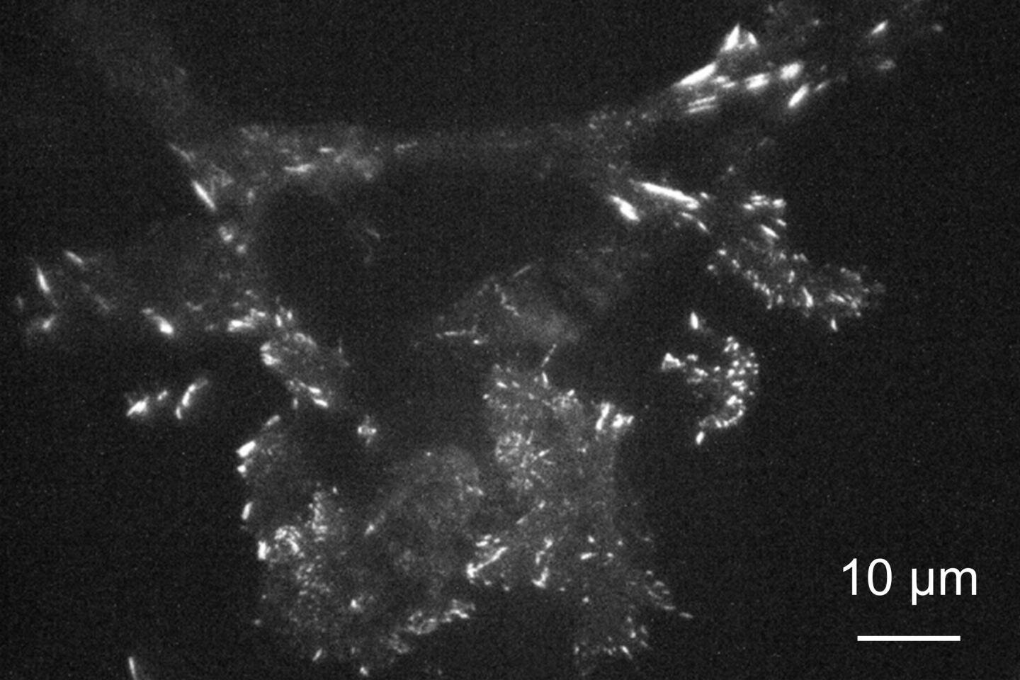
In the quest to image exceedingly small structures and phenomena with higher precision, scientists have been pushing the limits of optical microscope resolution, but these advances often come with increased complication and cost.
Now, researchers in Japan have shown that a glass surface embedded with self-assembled gold nanoparticles can improve resolution with little added cost even using a conventional widefield microscope, facilitating high-resolution fluorescence microscopy capable of high-speed imaging of living cells.
Because optical microscopes magnify light to obtain detailed images of a structure, the size of objects that can be distinguished has long been limited by diffraction—a property of light that causes it to spread when passing through an opening.
Researchers have been developing techniques to overcome these limits with highly advanced optical systems, but many of them depend on the use of strong lasers, which can damage or even kill living cells, and scanning of the sample or processing of multiple images, which inhibits real-time imaging.
“Recent techniques can produce stunning images, but many of them require highly specialized equipment and are incapable of observing the movement of living cells,” says Kaoru Tamada, distinguished professor at Kyushu University’s Institute for Materials Chemistry and Engineering.
Imaging cells using real-time fluorescence microscopy methods, Tamada and her group found that they could improve resolution under a conventional widefield microscope to near the diffraction limit just by changing the surface under the cells.
In fluorescence microscopy, cell structures of interest are tagged with molecules that absorb energy from incoming light and, through the process of fluorescence, re-emit it as light of a different color, which is collected to form the image.
Though cells are usually imaged on plain glass, Tamada’s group coated the glass surface with a self-assembled layer of gold nanoparticles covered with a thin layer of silicon dioxide, creating a so-called metasurface with special optical properties.
Only 12 nm in diameter, the organized metal nanoparticles exhibit a phenomenon known as localized surface plasmon resonance, which allows the metasurface to collect energy from nearby light-emitting molecules for highly efficient re-emission, thereby producing enhanced emission confined to the 10-nm thick nanoparticle surface.


