Zebrafish are widely used in developmental biology, immunology, neurobiology, and drug research, showing great progress (Advanced Zebrafish 3D Imaging System for Multidisciplinary Studies) in large-scale, multidisciplinary studies of drug development and effects in ‘because of its small size. However, effective experimental methods rely heavily on manual control and imaging under traditional microscopes with high performance but low performance, which limits the use of zebrafish in large-scale research.
To solve these problems, researchers from the Suzhou Institute of Biomedical Technology and Engineering (SIBET) and the Shanghai Institute of Nutrition and Health (SINH) of the Chinese Academy of Sciences (CAS) have developed a light-emitting diode system (LS- FIS) for three-dimensional (3D) visualization (Advanced Zebrafish 3D Imaging System for Multidisciplinary Studies) of zebrafish based on light sheets.
Light sheet microscopy is a 3D imaging system with low phototoxicity and fast imaging speed. However, current sheet imaging requires complex sample preparation procedures for zebrafish imaging. In addition, due to the limited field of view, 3D imaging of whole embryos often requires imaging and regional summation, which limits the imaging utility of this technique.
Using the intelligent water system and the optical connection system and the sample scheduling and the 3D algorithm reconstruction, the researchers combined the flow technology and the light of the sheet to obtain a high-quality 3D image of 200 embryos / hour.
LS-FIS has been applied to study the development of blood vessels in the trunk and head of zebrafish. 100 Tg (kdrl:EGFP) transgenic zebrafish, vascular endothelial cells labeled with green fluorescent protein were imaged daily with LS-FIS from three days to nine days post-fertilization for demonstration purposes.
More than 500 3D images of complete zebrafish embryos were obtained, which is the first known report of large-scale 3D imaging of an entire fish.
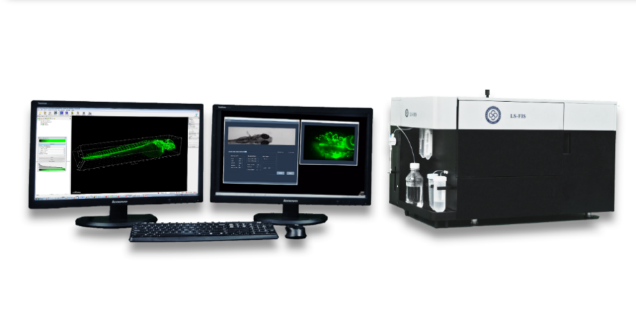
The researchers compared and analyzed the length of the central vessels and the changes in the shape of the vascular hyaloid in the 3D system obtained. The results show a significant diversity of the development of the vessels of the trunk but little heterogeneity of vascular hyaloid.
The work titled “Heterogeneities of Zebrafish Vascular Development Studied from a High Throughput Lightsheet Flow Imaging System” was published in Biomedical Optics Express.
Furthermore, the LS-FIS model was optimized for several iterations for better stability, better appearance and easier to use software interface. It has also been tested at SINH and CAS Institute of Neuroscience and found to have good imaging capabilities and usability. The researchers later developed image analysis and processing algorithms for processing large 3D image data captured by LS-FIS. For zebrafish intersegmental blood vessels, they proposed a multi-feature 3D convolutional neural network (MS-3D U-Net) to achieve 3D vessel segmentation and recognition. With multi-feature learning and optimal loss function based on dynamic estimation method, MS-3D U-net achieves accuracy of more than 90% (AUC value).
Based on MS-3D U-Net, they automatically segmented and measured 3D image data of zebrafish embryos observed anteriorly for 24 hours and plotted the development of lateral vessels and dorsal longitudinal vessels. anastomoses.


