Microrobots have been developed to connect neural networks to the environment in vivo. The research team of Professor Hongsoo Choi and Professor Yongseok Oh from DGIST joined the research team of Dr. Jongcheol Rah from the Korea Brain Research Institute is leading to develop (Can we connect to a virtual world like in the movie “Matrix”?) a technology to deliver a microrobot to the target area of the hippocampus in vitro. around, connect neural networks and measure neural signals
– Research results are expected to contribute to neural network research and validation and evaluation of cell therapy products
The research team of DGIST Professor Hongsoo Choi (Chairman Kuk Yang) of the Department of Robotics and Mechatronics Engineering has developed a microrobot capable of training neural networks in the hippocampal tissue section in an in vitro and ex vivo environment. Through joint research with Dr. Led by Jongcheol Rah from the Korea Brain Research Institute, it has been confirmed that neural networks can be analyzed structurally and functionally using a microrobot in an in vitro environment during cell administration and transplantation. The results of the research are expected to be applied in various areas, including neural networks, cell therapy products, and regenerative medicine.
Cell therapy products and cell delivery technologies have been developed to regenerate nerve cells damaged by disease; In recent years, various technologies related to microrobots that can deliver specific and small cells are being recognized more and more. Previous studies on cell transfer and neural network connectivity using microrobots have only investigated the structural and functional connectivity of cells at the cellular level.
The research team led by Professor Choi used microrobots that can be applied to neural networks in a practical way. This technology uses microrobots to enable the analysis of functionally connected neural networks in an ex vivo environment and cell delivery; Brain tissue from laboratory mice was used for the experiment.
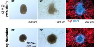
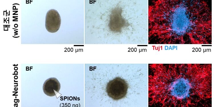
The research team first implanted super paramagnetic iron oxide nanoparticles into nerve cells in the hippocampus of laboratory mice to create the Mag-Neurobot into a three-dimensional shape. Magnetic nanoparticles are attached to the outside of the robot so that the robot can move to the desired location by reacting to the magnetic field. Safety was also verified through biocompatibility testing, in which the robot’s magnetism did not affect muscle cell growth.
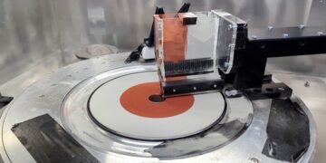
The research team implanted the microrobot into the tissue part of the hippocampus of a mouse using magnetic field control. Using immunofluorescence staining, the team found that the cells from the microrobot and the cells from the hippocampal tissue were neurites connected to each other in the structure.
In addition, a microelectrode array (MEA) was used to stimulate the microrobot nerve cells to determine whether the nerve cells delivered by the microrobot exhibit electrophysiological characteristics. It has been found that electrical signals are often propagated by nerve cells in the hippocampus. As a result, the research team confirmed that the neural cells generated by the microrobot can work effectively in cells and neural networks in the hippocampal tissue of laboratory mice. In addition, the team demonstrated that the microrobot can perform the task of delivering nerve cells and training neural networks.
Dr Choi from DGIST said: “We have shown that the microrobot and the muscle tissue from the mouse brain can integrate functions through electrophysiological analysis” and added: “The technology developed in this study should be used to identify targeted therapies in a precise manner in the field of neurology and cell therapy.
Source: Daegu Gyeongbuk Institute of Science & Technlogy ( DGIST).
