


The hidden structure behind a widely used class of materials
Relaxor ferroelectrics are materials used in electronics and sensors for a long time, but where their special properties came from was a mystery until now.
These materials have been used for many years in things like ultrasound, microphones, and sonar. Their special features are because of how their atoms are arranged, but people couldn’t directly see that arrangement until now.
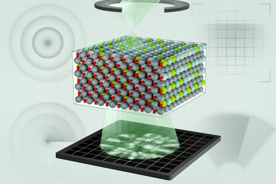
A group of researchers from MIT and other places have now shown the three-dimensional structure of atoms in a relaxor ferroelectric for the first time. Their findings, published in the journal Science, give a better way to model and design future technologies in computing, energy, and sensing.James LeBeau, who leads the research, says that now that they understand more about what’s happening, they can better predict and make materials with the properties they want.
“We are still learning how to engineer these materials,”he says. “But if we want to know if our models are right, we need to understand the real structure.
“In their study, the researchers used a new method to see how electric charges are spread in the material, which led to some surprising results.
Michael Xu and Menglin Zhu, who are both postdocs at MIT, say they realized the chemical mixing they saw in their experiments wasn’t fully considered before. By working with their team, they combined their findings with computer models to improve the models and make better predictions.
Other researchers on the paper include PhD students from MIT, professors from other universities, and researchers from different institutions.The study focused on a type of relaxor ferroelectric used in sensors and defense systems. It’s a mix of lead magnesium niobate and lead titanate.
The team used a new technique called multi-slice electron ptychography (MEP) to study it. This method uses a tiny probe of high-energy electrons to move across the material and measure how they scatter.
Zhu explains that the method works by taking multiple diffraction patterns and using them together to build a 3D picture. The technique revealed different levels of chemical and polar structures, from the atomic level to larger scales. The researchers also found that the areas with different polarizations were much smaller than what the existing models predicted.
They then used this new data to improve their computer models and make them more accurate. Xu says the old models just had random areas of polarization but didn’t show how they were connected.
Now they can show how individual atoms affect polarization based on their charge. Zhu thinks the paper shows how useful electron ptychography is for studying complex materials and offers new ways to study disordered materials.
Xu also says this is the first time they’ve linked the 3D structure of relaxor ferroelectrics to computer simulations in an electron microscope.
It proves that this technique can get detailed 3D information from a material. The researchers believe this method could one day help create new materials with better electronic properties for better memory, sensing, and energy technologies.
LeBeau says that materials science is becoming more complex as computers and AI get better. “If our models are not accurate, we can’t trust the results,” he says.
“This technique helps us understand why a material works the way it does and check if our models are correct.”
Source: Massachusetts Institute of Technology


