


A faster and more precise biomedical imaging technique discovered
MIT researchers discovered a surprising phenomenon in optical physics that could lead to a faster and more precise biomedical imaging technique.
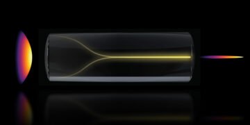
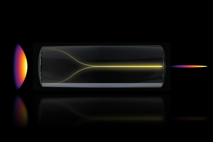
They found that under certain conditions, a chaotic mix of laser light can spontaneously organize into a highly focused “pencil beam”.
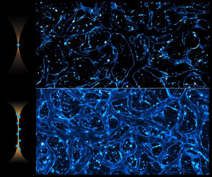
“Using this self-organized pencil beam, the team captured 3D images of the human blood-brain barrier 25 times faster than the standard method, while keeping the same level of detail.
This technology allows scientists to see individual cells absorbing drugs in real-time, which could help test new treatments for diseases like Alzheimers and ALS to see if they reach the right parts of the brain faster and more clearly.
Sixian You, an assistant professor at MIT, explained, “The common belief in the field was that increasing the laser power would make the light more chaotic. But we proved that’s not true. We followed the evidence, embraced the uncertainty, and found a way to let the light organize itself into a novel solution for bioimaging.”The discovery began with an observation that initially confused the researchers.
The team had developed a fiber shaper that allowed them to carefully adjust laser light passing through a multimode optical fiber. Cao was testing the fiber’s limits by increasing the power. Typically, more power makes the light more scattered, but Cao noticed that as he approached the fiber’s limit, the light instead collapsed into a single, sharp beam.
To replicate this effect, the researchers had to follow two specific conditions. First, the laser had to enter the fiber at a perfect angle, zero degrees. Second, the power had to be increased until the light started interacting with the fiber’s glass.
“Once the laser reaches this critical power, the nonlinearity balances the fiber’s natural disorder, creating a stable, ultrafast pencil beam,” Cao explained.
Most researchers avoid high power levels to prevent damaging the fiber, and precise alignment isn’t often necessary because multimode fibers can carry so much power. However, together, these techniques can produce a stable pencil beam without complex setup.
“This method is neat because you can use a normal optical setup without needing deep expertise,” You said.The pencil beam proved more stable and high-resolution than other beams, which often have “sidelobes” blurry halos that can mess up images. Their beam was clean and sharply focused.
Using this pencil beam, the researchers demonstrated its use in imaging the human blood-brain barrier, a tightly packed layer of cells that protects the brain but also blocks many medicines. Scientists and doctors want to see how drugs move through this barrier and reach their targets in the brain.
With standard methods, researchers can only capture one 2D section at a time and repeat the process multiple times. The new technique allows real-time tracking of how cells absorb proteins, creating ultrafast, high-precision images.
Roger Kamm, a professor at MIT, said, “The pharmaceutical industry is really interested in using human-based models to find drugs that can cross the barrier, as animal models often don’t predict what happens in humans. That this new method doesn’t require fluorescent tags is a game-changer.
For the first time, we can now visualize the time-dependent entry of drugs into the brain and even measure how fast different cell types take in the drug.”
“Importantly, however, this method isn’t just for the blood-brain barrier. It allows for tracking different compounds and molecules across various tissue models over time, making it a strong tool for biological engineering,” Spitz says.
The team created 3D images of cells that were better quality than with other methods, and they produced these images about 25 times faster.
“Normally, there’s a tradeoff between image clarity and how deep you can look at once — you can’t go too deep at the same time.
But with our method, we can beat this tradeoff by creating a pencil-beam that has both high clarity and a wide depth of focus,” You explains.
Looking ahead, the researchers want to better understand the basic physics behind the pencil-beam and how it self-organizes.
They also plan to use the technique in other situations, like imaging brain neurons, and work toward making the technology available for commercial use.
“You’s group created a beam that focuses energy in both time and space, which could be useful for microscopy that relies on light intensity.
They showed this and found it better than regular laser beams for imaging. It would be interesting to fully understand how these new pencil beams are made, as they could be useful in many imaging applications,” says Frank Wise, the Samuel B. Eckert Professor of Engineering Emeritus at Cornell University, who wasn’t involved in this work.
Source: Massachusetts Institute of Technology


